“What will the wound look like?” is an important question before Mohs surgery. The repair is tailored to the treated area and the wound left after the tumour has been removed. Understanding the options helps you discuss them with your surgeon without assuming that everyone needs the same technique.
Reconstruction after Mohs surgery: how is the skin repaired after skin cancer removal?
Mohs surgery is one of the most precise and effective techniques for treating certain skin cancers, particularly basal cell carcinoma and squamous cell carcinoma. Its main advantage is that it allows the tumour to be removed with microscopic control of the surgical margins while preserving as much healthy tissue as possible.
Once the cancer has been removed, however, patients often ask: how will the wound left by surgery be closed?
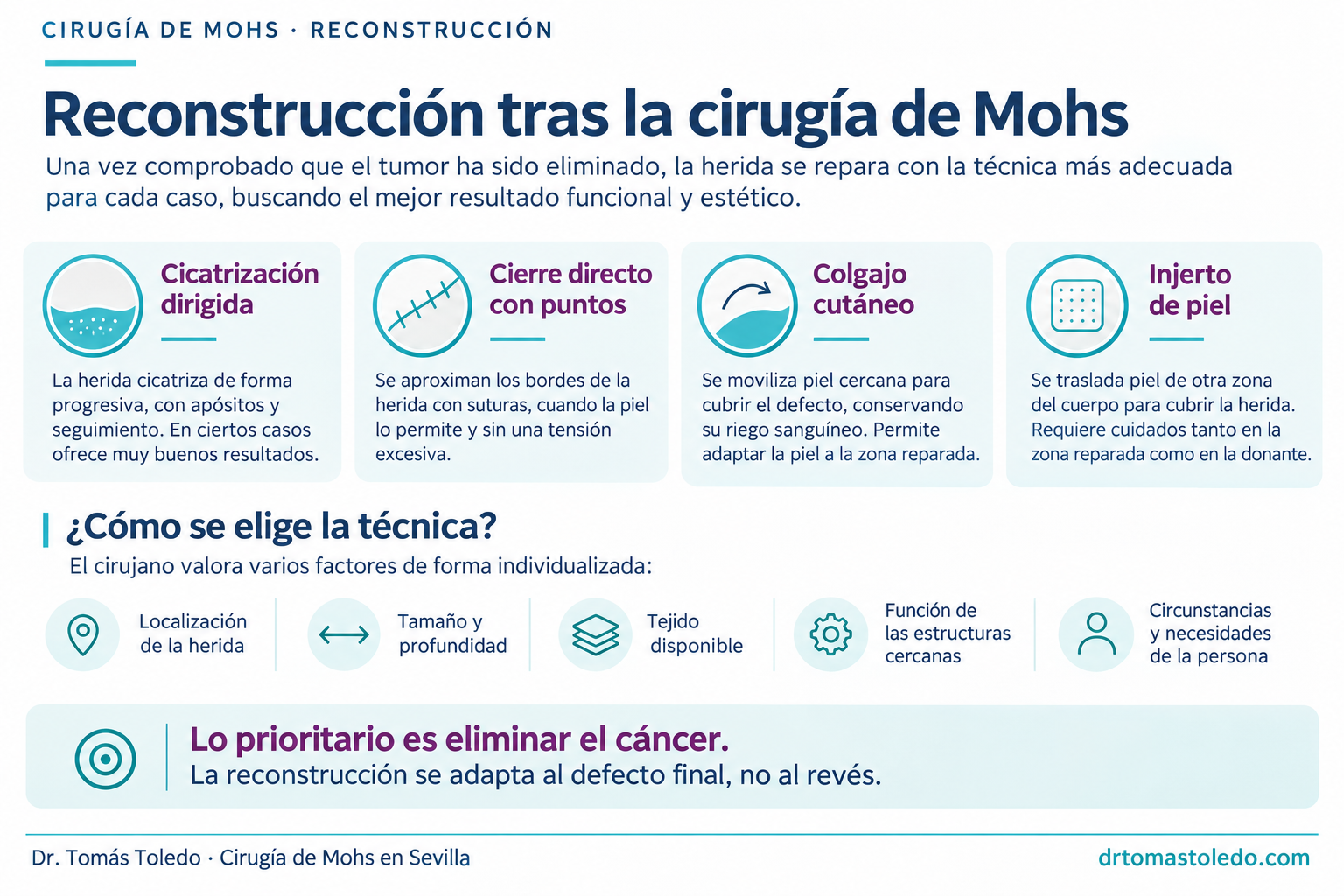
The answer depends on several factors. Some wounds can be closed directly with stitches, others require nearby skin to be moved using a flap or a skin graft, and in selected cases the best option may be to allow the wound to heal gradually.
Although reconstruction is a fundamental part of treatment, one essential principle should be remembered: Mohs surgery is first and foremost cancer surgery. Its priority is to remove the tumour completely. Reconstruction is planned afterwards, seeking the best possible balance between safety, function and cosmetic outcome.
First remove the cancer, then reconstruct
One of the main differences between Mohs surgery and conventional skin cancer surgery is how the removal of the tumour is checked.
In conventional surgery, the lesion is usually removed with a margin of apparently healthy skin and the specimen is sent to the pathology laboratory. The final result is generally available several days after the operation.
In Mohs micrographic surgery, the removed tissue is examined microscopically using a technique that systematically assesses the peripheral and deep surgical margins.
If tumour cells are found at a margin, the surgeon can remove another layer only from the affected area. This process is repeated until the examined margins are free of tumour.
This margin control has two major advantages: it allows high cure rates and avoids unnecessary removal of healthy tissue.
Preserving tissue is especially important when the cancer is in a delicate area such as the nose, eyelids, lips or ears, where even a small loss of skin can have functional or cosmetic consequences.
Definitive reconstruction is considered once the tumour has been removed and margin clearance confirmed.
In conventional fresh-tissue Mohs surgery, reconstruction usually takes place on the same day. In some circumstances, it may be preferable to perform it later, particularly if more complex planning or the involvement of other specialists is needed.
This approach is also reflected in multidisciplinary recommendations on reconstruction after skin cancer removal, developed with the participation of organisations such as the American Academy of Dermatology and the American College of Mohs Surgery.
Who performs reconstruction after Mohs surgery?
A common question is whether reconstruction must be performed by a plastic surgeon.
The answer is no. A dermatological surgeon specialising in Mohs surgery receives specific training both in removing skin tumours under microscopic control and in reconstructing the resulting surgical defects.
Reconstruction is part of a Mohs surgeon’s routine practice and ranges from direct closure to considerably complex skin flaps and grafts.
Having the same specialist perform tumour removal and reconstruction can offer important advantages. The surgeon knows the exact location, depth and extent of the defect, as well as the characteristics of the surrounding tissue.
This allows reconstructive planning to be integrated into the overall surgical strategy.
In selected cases, especially with very extensive defects or involvement of deeper anatomical structures, collaboration with other specialists may be appropriate, including plastic surgeons, ophthalmologists specialising in oculoplastic surgery or ear, nose and throat surgeons.
This collaboration does not mean that routine reconstruction must be performed by another specialist. Each procedure should be adapted to the patient’s particular needs.
The four main options for reconstruction after Mohs
There is no single reconstructive technique suitable for every wound.
The aim is to choose the technique that offers the best possible outcome with the lowest risk and the least disturbance to surrounding tissue.
1. Healing by secondary intention
Although many people associate an operation with stitches, some wounds can heal well without sutures.
This approach is called healing by secondary intention, or guided wound healing.
It involves allowing the body to repair the wound gradually by forming granulation tissue and then covering the surface with new skin.
During this process, the wound becomes smaller and gradually takes on an appearance closer to that of the surrounding skin.
Healing by secondary intention can be a deliberate reconstructive choice and, in selected cases, provide excellent cosmetic and functional results.
It can be particularly useful on certain concave surfaces, such as parts of the ear or nose, where the anatomy itself can favour satisfactory healing.
Its main advantages are that it avoids additional incisions, does not require a skin donor site and leaves the surrounding tissues intact.
However, it also has limitations.
Healing is usually slower than after closure with stitches and requires regular dressing changes, wound protection and follow-up.
The natural contraction of tissue during healing can also cause distortion when a wound is close to moving structures or free edges, such as the eyelids or lips.
Not every location is therefore suitable for this approach.
Allowing a wound to heal does not mean leaving it without care. It is a reconstructive technique that requires appropriate selection and specific aftercare.
2. Direct closure with sutures
Direct closure is probably the most familiar form of reconstruction.
It involves bringing the wound edges together and holding them with stitches so that the tissues heal in the intended position.
It may appear to be the simplest technique, but good closure requires several factors to be assessed.
The surgeon must check that the skin is sufficiently mobile, that the edges can be brought together without excessive tension, and that closure will not displace important anatomical structures.
Sometimes the incision needs to be made slightly longer so that the edges fit together correctly.
This explains something that can surprise patients: after removal of an apparently small tumour, the resulting scar may be considerably longer than the original lesion’s diameter.
This does not mean that unnecessary tissue has been removed. In many cases, this adjustment creates a better-oriented linear scar and distributes tension appropriately.
Where possible, the surgeon places the scar along natural skin tension lines or anatomical folds so that it becomes less noticeable over time.
In other cases, internal stitches provide support for deeper tissues and reduce tension on the superficial sutures.
Direct closure can offer a relatively quick recovery and a very satisfactory result, but it is not always the best option.
Closure under excessive tension can impair healing, widen the scar or alter the position of nearby structures.
For this reason, a wound should not be closed directly simply because its edges can technically be brought together.
3. Reconstruction with skin flaps
When an appropriate direct closure is not possible, reconstruction with a flap is one of the most commonly used alternatives.
A flap involves moving skin and sometimes subcutaneous tissue from a nearby area to cover the defect left by cancer removal.
Its defining feature is that the moved tissue retains a vascular connection to its original location, preserving its blood supply.
There are many types of flap, including advancement, rotation and transposition flaps.
Each design moves skin in a different way to suit the shape of the defect and the characteristics of the anatomical region.
For example, a defect on the nose may be repaired using nearby skin with similar thickness, texture and colour.
The main advantage of nearby tissue is that it usually closely resembles the skin that has been removed.
This is particularly important on the face, where differences in texture, pigmentation and thickness can be very visible.
Flaps also redistribute tension and can prevent healing from distorting delicate structures.
Their design nevertheless requires precise planning.
A poorly oriented flap can change the natural contours of the face, alter an eyelid’s position or cause nasal asymmetry.
Additional incisions may be needed, creating scars outside the area initially treated.
Although these incisions may look striking in a diagram, they are usually positioned to fit within the skin’s natural lines and folds.
Flaps are a very useful reconstructive tool, but they should be selected to suit the individual defect rather than for their technical complexity.
4. Reconstruction with skin grafts
Another option is a skin graft.
Unlike a flap, which retains its original vascular connection, a graft involves taking a piece of skin from another part of the body and transferring it completely to the area requiring repair.
Once in place, the tissue must gradually establish a new blood supply from the recipient wound bed to survive.
Dermatological surgery frequently uses full-thickness skin grafts, which contain the epidermis and the entire dermis.
Depending on the defect, skin may be taken from different areas, such as in front of or behind the ear or above the collarbone.
The donor site is chosen to match the characteristics of the area being reconstructed as closely as possible.
Grafts can be particularly useful when there is insufficient nearby skin for direct closure or when moving adjacent tissue would cause unnecessary distortion.
They also allow some defects to be repaired without displacing nearby anatomical structures.
However, they have particular considerations.
First, two areas must heal: the recipient site where the graft is placed and the donor site from which the skin is taken.
Second, during the first few days it is essential to avoid movement, inappropriate pressure or collections of blood that could interfere with graft integration.
Special securing dressings are sometimes used to immobilise the graft while its blood supply develops.
Even when donor skin is carefully selected, there may be small differences in colour, texture or contour compared with the surrounding skin.
In some cases these differences gradually improve; in others they may partly persist.
The choice of a graft must therefore consider both whether it can cover the defect and the cosmetic and functional characteristics of the area.
How does the surgeon choose the most suitable reconstruction?
The choice of reconstructive technique does not depend on wound size alone.
Two surgical defects of similar dimensions may require completely different procedures depending on their location and depth.
The surgeon pays particular attention to the following factors.
Anatomical location
Skin does not have the same characteristics throughout the body.
The cheek usually has more skin mobility than the nose. In the eyelids, small changes in tension can have important functional consequences.
A technique suitable for the scalp may be entirely inappropriate for the upper lip.
Wound depth
Repairing a superficial loss of skin is different from repairing a defect that extends into fat, muscle, cartilage or other deep structures.
Depth determines which tissues need repair and which techniques can be used safely.
Mobility and quality of the surrounding skin
Elasticity, thickness, previous scars and sun damage influence the reconstructive options.
Previous operations or radiotherapy in the area also affect the choice.
Personal and medical factors
Age, general health, certain illnesses, smoking and some treatments can influence healing.
Functional needs, the ability to manage wound care and the patient’s preferences must also be considered.
For example, someone who has difficulty attending frequent reviews may benefit from a different strategy from someone able to undertake prolonged wound care more easily.
Expected functional and cosmetic outcome
Reconstruction aims to restore anatomy and preserve the normal function of the area.
Appearance matters, but it must never compromise treatment safety or the function of the structures involved.
Can the necessary reconstruction be predicted before surgery?
This is one of the most common questions during the preoperative consultation.
For many patients, the most likely reconstructive options can be anticipated.
However, the final technique cannot always be determined before complete tumour removal.
The reason is straightforward: the microscopic extent of a skin cancer does not always match its visible size.
Some tumours have microscopic extensions into apparently healthy skin. This is particularly relevant to certain infiltrative basal cell carcinomas and lesions that have returned after previous treatment.
Several stages of removal may be needed during Mohs surgery to achieve clear margins.
As a result, the final defect may be larger or differently shaped than initially expected.
Although the surgeon can explain the reconstructive possibilities during the consultation, the final decision must be made once the true extent of the wound is known.
This reflects the priority of treating the cancer completely and then adapting the repair to the actual needs of the wound.
A final consideration: Mohs success is about more than the scar
Mohs micrographic surgery combines two complementary aims: controlling the cancer and preserving as much healthy tissue as possible.
Reconstruction is an essential part of that process, especially in areas where small anatomical changes can have significant functional and cosmetic effects.
It is nevertheless important to keep the main purpose of the operation in mind.
This is cancer surgery. The primary aim is to remove the cancer as thoroughly as possible.
Once that aim has been achieved, reconstruction must be tailored to the actual defect, the anatomy and the patient’s circumstances.
The most complex operation is not always needed. Sometimes well-placed stitches or healing by secondary intention provide the best solution. In other cases, flaps or grafts are essential to restore anatomy and function.
Combining microscopic margin control, knowledge of anatomy and experience in reconstructive surgery allows both aims to be addressed together.
Good Mohs surgery involves treating the cancer appropriately and restoring the best possible function and appearance to the treated area without compromising cancer treatment safety. Follow-up remains important after wound repair.
Sources
Original article by Dr Tomás Toledo.
AAD: guideline on reconstruction after skin cancer resection.
AAD: basal cell carcinoma and repair options.
British Association of Dermatologists: Mohs surgery.
Cambridge University Hospitals: repair and risks of Mohs surgery.
General information for patients. This does not replace instructions from Dr Tomás Toledo’s team.